Doppler ultrasound and elastography

Doppler ultrasound and elastography

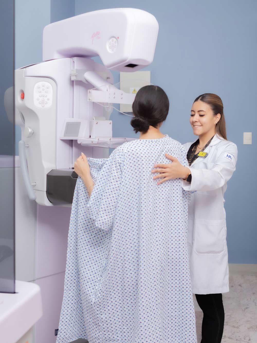
Doppler ultrasound and elastography is a non-invasive test that combines advanced techniques to accurately evaluate organs and tissues. We use state-of-the-art technology and have trained professionals to provide you with reliable diagnoses.


White monitor displaying a Doppler ultrasound White monitor displaying a Doppler ultrasound

Blue location pin icon on a white background. 909 Paseo del Tecnológico, Residencial Tecnológico, 27250 Torreón, Coahuila, México

+52 871 729 0400 Ext. 416

24 hours a day, 365 days a year

The Doppler ultrasound and elastography service at Hospital Angeles Torreón two non-invasive imaging techniques to provide timely and accurate diagnoses across multiple medical specialties. Doppler ultrasound allows for real-time observation of blood flow and assessment of arteries and veins, while elastography evaluates tissue stiffness and elasticity to aid in the early detection of fibrosis, inflammation, or masses. This combination, also known as Doppler ultrasound with elastography, does not use ionizing radiation, making it safe for various patient groups and useful as a guide during procedures, including ultrasound-guided biopsies when indicated by your doctor.

In the cardiovascular context, Doppler ultrasound helps identify flow abnormalities associated with peripheral arterial or venous disease and provides complementary data for the evaluation of the heart and great vessels. In organs such as the liver, kidney, and thyroid, elastography adds information about tissue consistency, which can guide the investigation of liver diseases or thyroid nodules and support the differentiation between benign and suspicious changes. The synergy between these two modalities improves comprehensive evaluation, expands the scope of conventional ultrasonography, and contributes to more accurate clinical decisions at Hospital Angeles Torreón.

Common indications include the analysis of circulation in the extremities, the monitoring of heart disease, and the assessment of solid organs. In hepatology, for example, elastography complements the clinical and laboratory profile to stratify liver stiffness, while Doppler provides data on vascular patterns. In thyroid and kidney imaging, these tools help characterize lesions using a functional and tissue-based approach. All of this is integrated into the clinical practice of the Department of Imaging, supported by advanced technology available at our facility and aligned with institutional standards.

With Doppler ultrasound and elastography at Hospital Angeles , you will receive a reliable, safe, and tailored evaluation for your diagnosis.

Frequently Asked Questions

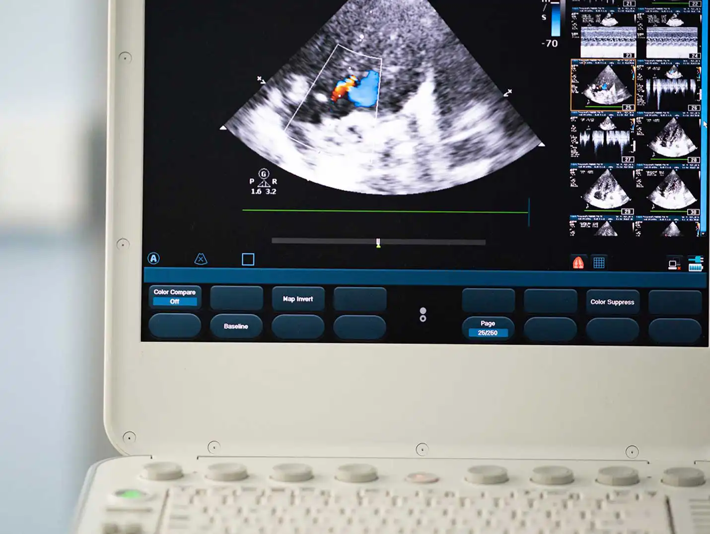
Doppler ultrasound and elastography is an imaging technique that uses sound waves to create images of the body's organs and tissues. It allows for the assessment of blood flow and tissue elasticity.

Doppler ultrasound and elastography are used to diagnose and monitor cardiovascular diseases, such as peripheral artery disease and heart disease. They are also used to assess liver, kidney, and thyroid function, as well as to detect tumors and evaluate tissue elasticity.

No, Doppler ultrasound and elastography are noninvasive and generally painless procedures. A conductive gel is applied to the skin, and a transducer is moved over the area to be examined to obtain the images.

The duration of a Doppler ultrasound and elastography can vary depending on the area being examined and the complexity of the case. Generally, the procedure takes between 30 and 60 minutes.

In some cases, special preparation may be required before a Doppler ultrasound and elastography. This may include fasting beforehand, avoiding certain medications or liquids, or following specific preparation instructions as directed by your doctor. It is important to follow the instructions provided by the medical staff.

Hospitals that offer this service

An icon of a list inside a blue circle. Next to it is a white box with the word "contact"