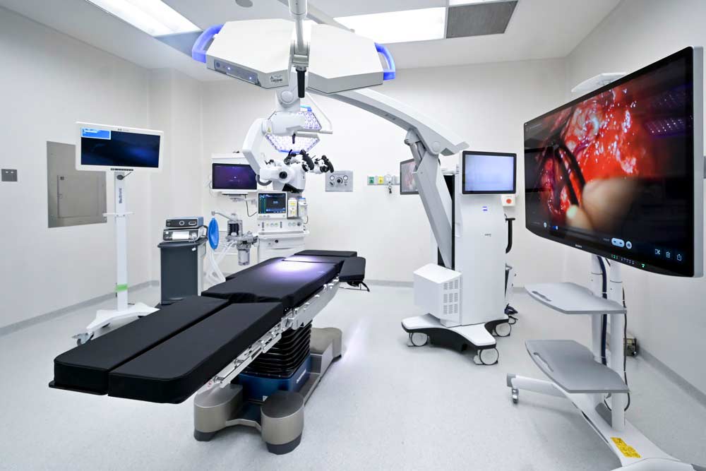
Imaging studies that aid in the diagnosis of diseases and guide surgical or therapeutic procedures, with the goal of providing the right treatment for each patient. The facility features advanced technology and an excellent medical staff.
The Department of Imaging is responsible for performing the necessary medical imaging studies. It was previously called Radiology (based on the fact that X-rays were the only primary source for obtaining images), but now other technologies that do not use X-rays (ultrasound and magnetic resonance imaging) have been incorporated. That is why it is called imaging.
The studies can be divided into categories based on the technology used to acquire theimages:
GENERAL RADIOLOGY AND FLUOROSCOPY
This includes “classic radiology” studies such as chest X-rays and X-rays of bones and joints. In most cases, contrast media can be used to evaluate various organs and systems (esophagus, stomach, and duodenum; excretory urography).
It uses ionizing radiation, and many of these tests require some preparation as well as scheduling an appointment.
ULTRASOUND
It uses sound waves, similar to sonar. It does not use radiation. Its use is becoming increasingly widespread and is almost always used to evaluate solid structures in the body (for example, the liver, the kidneys). Structures containing fluid (the gallbladder when fasting, the full urinary bladder) can also be studied in great detail.
There are different probes (transducers) used to evaluate multiple areas of the body (liver, gallbladder, uterus, ovaries, thyroid, breast, and prostate). Some involve scanning the body’s surface, while others can be performed through body cavities (rectally or transvaginally).
There is also a technique called Doppler, which is used to examine vascular structures (arteries and/or veins) using non-invasive methods. Many exams require some form of preparation and a prior appointment.
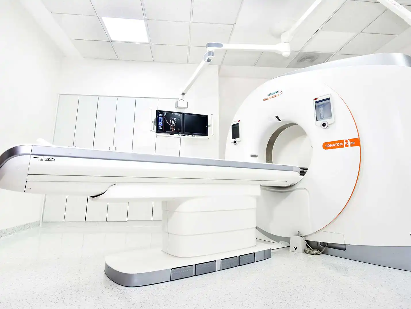
COMPUTED TOMOGRAPHY
MAMMOGRAPHY (MASTOMOGRAPHY)
This imaging modality uses radiation (X-rays) to provide a comprehensive assessment of the breast. Our equipment is among the most advanced in the world; it is fully digital and integrated with a computer-aided detection (CAD) program.
This procedure involves placing the breast on a surface, applying brief direct compression, and taking an X-ray. Typically, 2 to 3 views are taken of each breast.
For patients with cosmetic implants (prostheses), at least 4 views must be taken of each breast.
This is currently the recommended method for screening or early detection of cancer; it is recommended starting at age 40 and should be performed annually. It requires preparation as well as a prior appointment.

 560 México Highway, Pastoresa, 91193 Xalapa, Ver.
  +52 228 141 0800
  7:00 a.m. to 9:00 p.m.