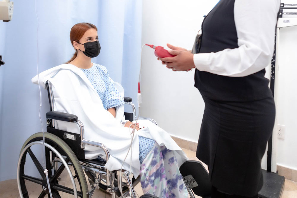
Cardiovascular Center / Cardiac Unit
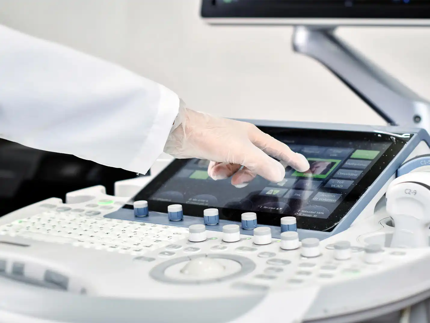
The echocardiography unit at Hospital Angeles cardiac examinations using high-resolution ultrasound and color Doppler imaging. It offers accurate, non-invasive echocardiograms tailored to each patient, utilizing advanced technology and specialists in cardiac imaging to assess heart health and function.
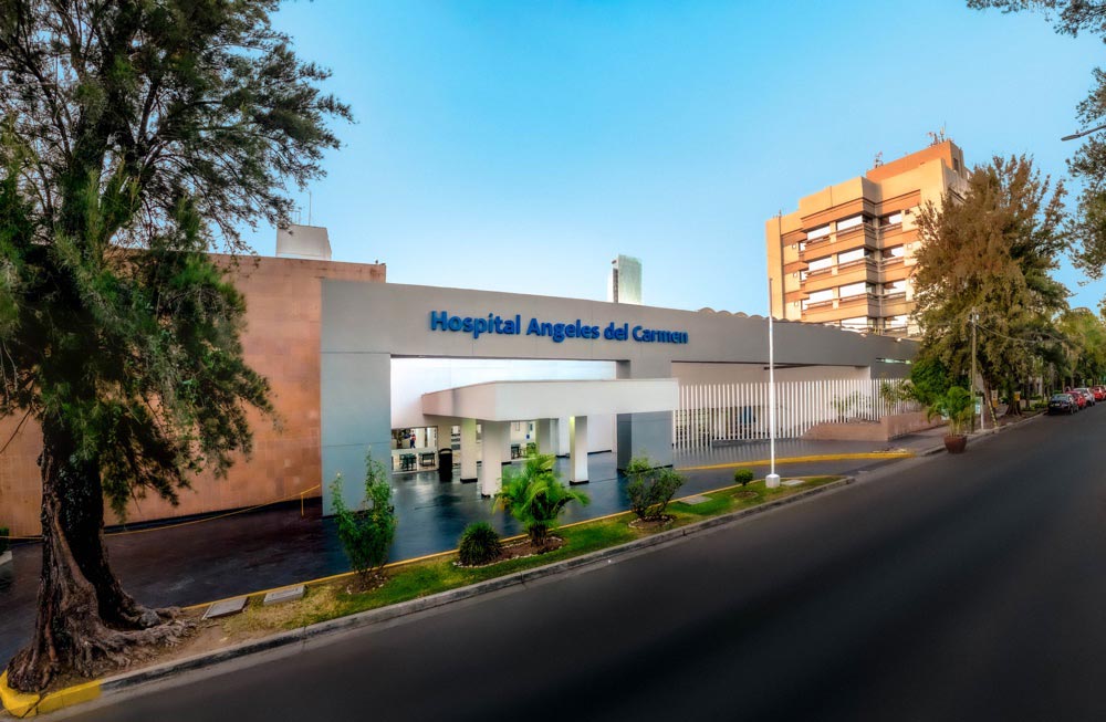
 3435 Tarascos Street, Monraz, 44670 Guadalajara, Jalisco
 +52 33 3813 0042
Ext. 2262
+52 33 3813 0042
Ext. 2262
 Monday through Friday, 9:00 a.m. to 6:00 p.m.
Echocardiography is a diagnostic test that uses ultrasound to produce images of the heart and assess its function. It is used to detect and evaluate heart conditions, such as heart valve problems, heart muscle disease, and congenital heart defects.
The cost of an echocardiogram at the Echocardiography Unit may vary depending on the type of test required and each patient’s specific circumstances. We recommend contacting the hospital directly for accurate information on costs.
In most cases, no special preparation is required for an echocardiogram. However, in certain cases, the doctor may instruct the patient to avoid eating or drinking for a period of time before the test.
The duration of an echocardiogram can vary depending on the condition and the individual characteristics of each patient. In general, the procedure usually takes between 30 and 60 minutes.
No, an echocardiogram is not painful. During the procedure, a conductive gel is applied to the patient’s chest, and an ultrasound transducer is used to obtain images of the heart. The patient may feel slight pressure on the chest due to the weight of the transducer, but should not experience any pain.