Echocardiography Unit

Echocardiography Unit

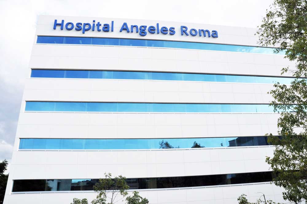
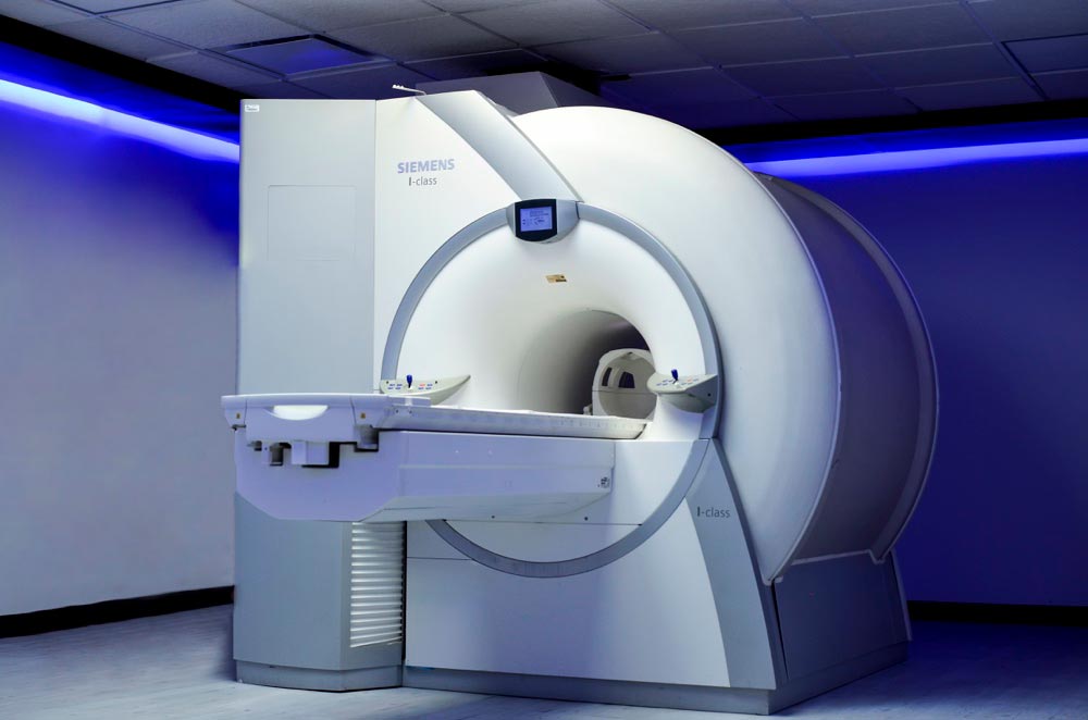
Cardiovascular Center / Cardiac Unit

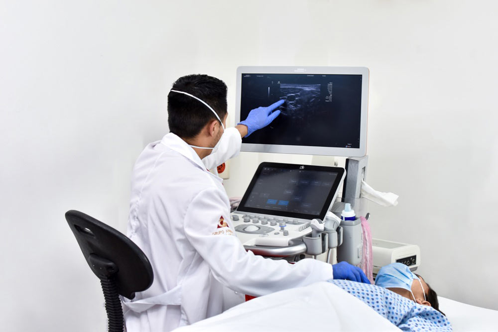
The echocardiography unit at Hospital Angeles cardiac examinations using high-resolution ultrasound and color Doppler imaging. It offers accurate, non-invasive echocardiograms tailored to each patient, utilizing advanced technology and specialists in cardiac imaging to assess heart health and function.


A doctor wearing a white coat and clear gloves pressing a button on a Voluson ultrasound machine. A doctor wearing a white coat and clear gloves pressing a button on a Voluson ultrasound machine.

Blue location pin icon on a white background. Querétaro Street, Roma ., 06700 México City, CDMX

+52 55 5265 3000 Ext. 2115

9:00 a.m. to 7:00 p.m.

The Echocardiography Unit at Hospital Angeles Roma specialized services for the diagnosis and monitoring of heart disease. Our team of cardiology experts uses state-of-the-art technology to perform high-quality echocardiographic studies.

Echocardiography is a non-invasive technique that uses ultrasound to obtain detailed images of the heart and its structures. This test allows us to assess heart function, detect abnormalities in the valves and blood vessels, and diagnose conditions such as heart failure, arrhythmias, and congenital defects.

At the Echocardiography Unit at Hospital Angeles Roma state-of-the-art equipment and a highly trained medical team to perform these studies accurately and reliably. Our goal is to provide our patients with an accurate and timely diagnosis so they can receive the appropriate treatment and improve their quality of life.

In addition to conventional echocardiography, we also offer specialized services such as transesophageal echocardiography and Doppler studies, which provide additional information about blood flow and heart function.

The Echocardiography Unit at Hospital Angeles Roma comprehensive, high-quality services for the diagnosis and monitoring of heart disease. Trust our team of experts and the cutting-edge technology we use to deliver accurate and reliable results.

Frequently Asked Questions

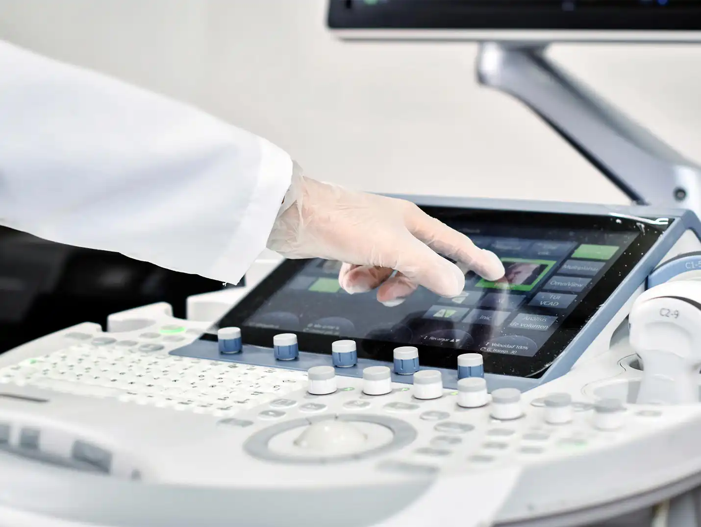
Echocardiography is a diagnostic test that uses ultrasound to produce images of the heart and assess its function. It is used to detect and evaluate heart conditions such as heart valve problems, heart muscle disease, and congenital defects.

During an echocardiogram, gel is applied to the chest and a transducer is placed on the skin. The transducer emits ultrasound waves that bounce off the heart and create real-time images. The doctor analyzes these images to assess the heart’s structure and function.

No, an echocardiogram is a painless procedure. The gel used may feel cold at first, but it does not cause any discomfort. The transducer is moved gently over the skin and does not cause any pain.

In most cases, no special preparation is needed before an echocardiogram. However, you may be asked to avoid eating or drinking for a certain period of time before the test, especially if you are having a transesophageal echocardiogram.

The duration of an echocardiogram can vary, but it generally takes between 30 and 60 minutes. The exact time will depend on the type of echocardiogram being performed and the information the doctor needs to obtain.

Hospitals that offer this service

An icon of a list inside a blue circle. Next to it is a white box with the word "contact"