It is a high-quality imaging method that has revolutionized medical diagnosis by providing detailed images of the body’s interior. Since it does not use ionizing radiation, such as X-rays, it is safe for patients of all ages and poses no risk of radiation exposure. As a result, this tool helps create accurate images of various parts of the body, including the brain, spine, joints, and internal organs.
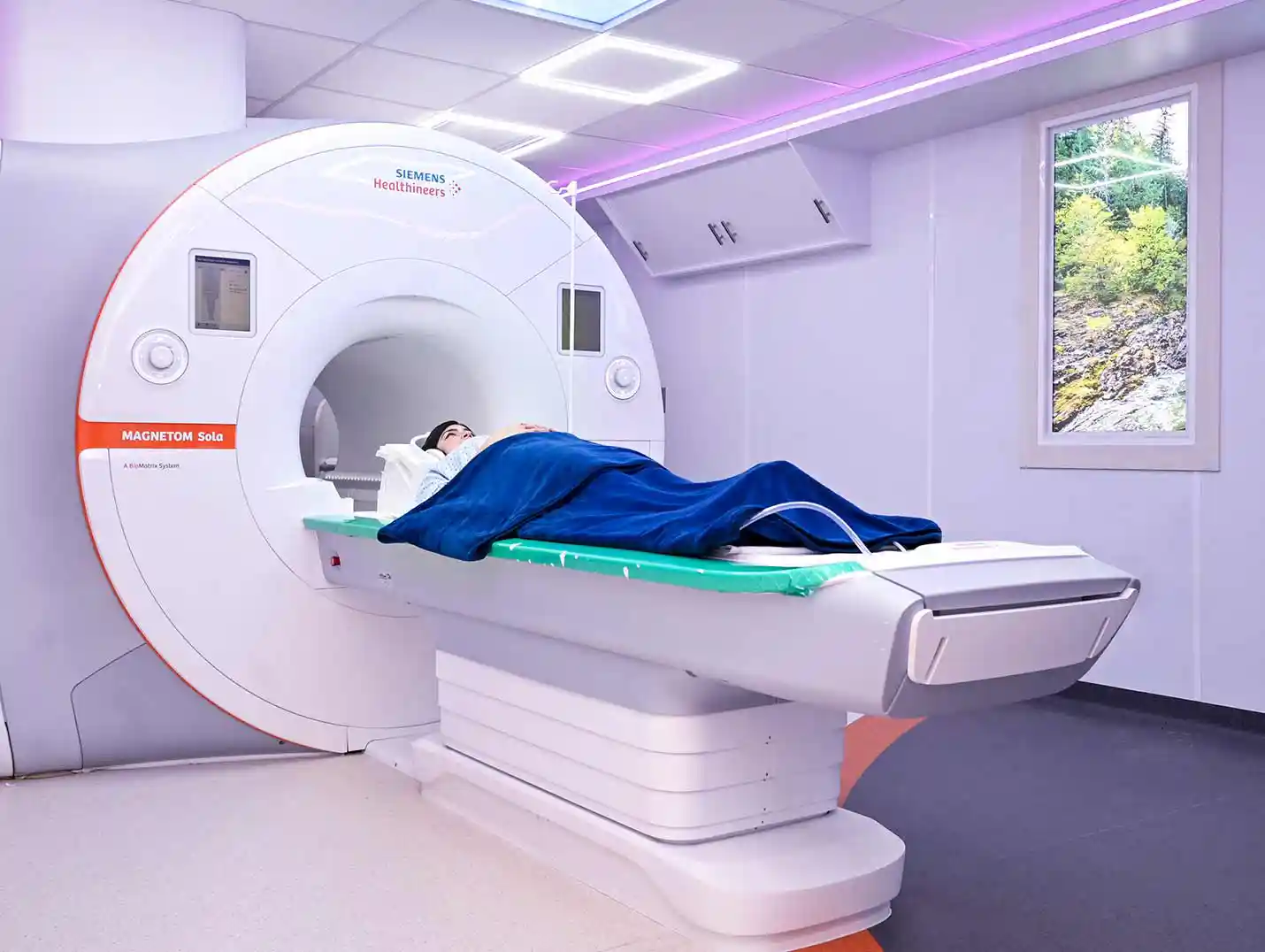
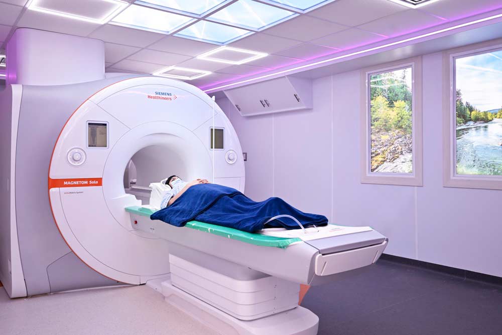
Magnetic resonance imaging (MRI) is a diagnostic imaging technique that uses magnets and radio waves to produce detailed images of the inside of the body. It works by generating a magnetic field that aligns the hydrogen atoms in the body, and then emitting radio waves to detect the signals emitted by these atoms and create images.
Magnetic resonance imaging (MRI) offers numerous benefits, as it provides detailed images of the body’s soft tissues, organs, and internal structures. It is particularly useful for diagnosing diseases and injuries in the brain, spine, joints, abdomen, and chest. Furthermore, it does not use ionizing radiation, making it safe and noninvasive.
Before an MRI, it is important to tell your doctor if you have any metal implants in your body, such as pacemakers, aneurysm clips, or joint replacements. In addition, you may need to avoid eating or drinking for a certain period of time before the exam, depending on the part of the body being examined.
An MRI scan itself is not painful. However, you may need to lie still during the exam, which some people find uncomfortable. In addition, you may be given an intravenous contrast dye to improve the quality of the images, which can cause a warm sensation in your body.
The duration of an MRI scan can vary depending on the part of the body being examined and the complexity of the case. Generally, the exam lasts between 30 and 60 minutes. However, it may take longer if additional images are needed or if an intravenous contrast agent is used.

 Av. Cerro Gordo, Lomas Campestre, 37150 León, Guanajuato, México
  +52 477 788 5600