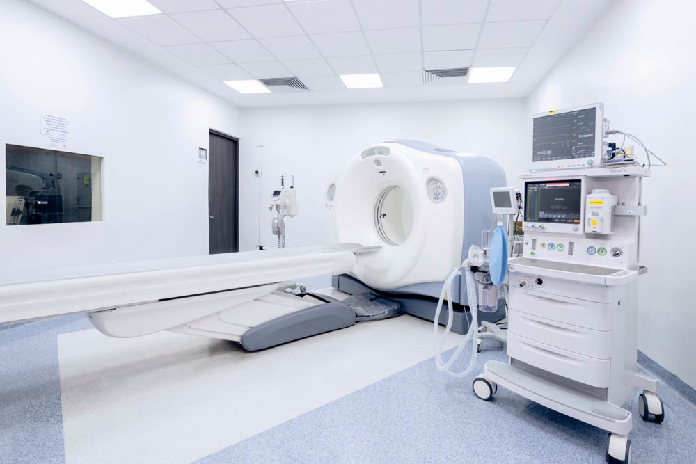
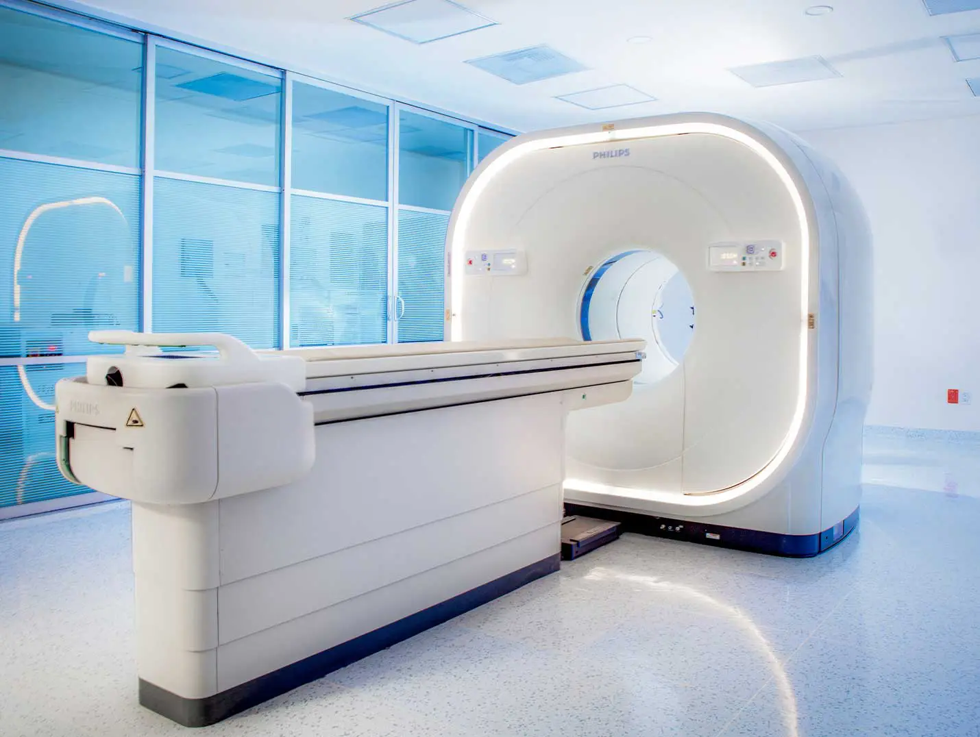
Positron emission tomography (PET) is a nuclear medicine test that combines metabolic and anatomical imaging in a single scan. At the Hospital Angeles PET service, Hospital Angeles standardized protocols and advanced technology to locate lesions, measure their biological activity, and correlate it with bodily structures. This technique is essential in oncology, as it allows us to assess disease spread, evaluate response to treatment, and plan therapies with greater precision—always with safety and specialized care.
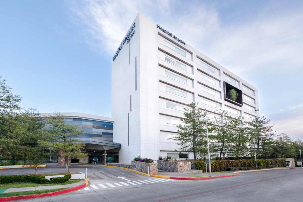
 331 Montaña Monarca Ave., North, Montaña Monarca Development, 58350 Morelia, Mich.
 +52 443 147 7150
Ext. 2219
+52 443 147 7150
Ext. 2219
 7:00 a.m. to 7:30 p.m.
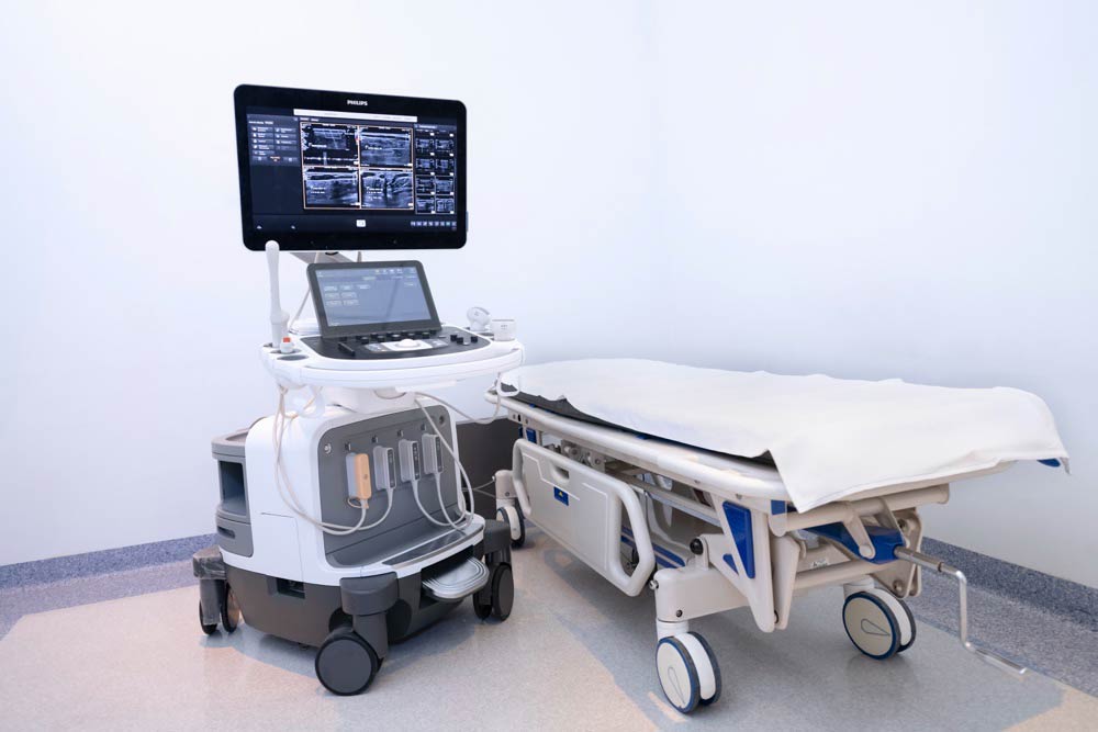
Positron emission tomography (PET-CT) is an imaging technique that combines computed tomography (CT) and positron emission tomography (PET) to produce detailed images of the inside of the body.
Positron emission tomography (PET-CT) is primarily used to detect and evaluate conditions such as cancer, heart disease, and neurological disorders.
During the procedure, the patient is injected with a small amount of a radioactive substance called a tracer. A series of images is then taken using a machine that combines computed tomography (CT) and positron emission tomography (PET).
Yes, positron emission tomography (PET-CT) is a safe procedure. However, because it uses radiation, there is a small risk of radiation exposure. The benefits of the procedure usually outweigh the risks.
Yes, before a positron emission tomography (PET-CT) scan, you must follow certain preparation instructions. These may include fasting beforehand, stopping certain medications, and avoiding caffeine. It is important to follow the instructions provided by your doctor or the diagnostic center.