Positron emission tomography (PET) is a nuclear medicine test that combines metabolic and anatomical imaging in a single scan. At the Hospital Angeles PET service, Hospital Angeles standardized protocols and advanced technology to locate lesions, measure their biological activity, and correlate it with bodily structures. This technique is essential in oncology, as it allows us to assess disease spread, evaluate response to treatment, and plan therapies with greater precision—always with safety and specialized care.
 1055-S Camino de Santa Teresa, Héroes de Padierna, 10700 México City.
 +52 55 5449 5500
Ext. 3043–3059
+52 55 5449 5500
Ext. 3043–3059
 By appointment
The Positron Emission Tomography (PET) Department at Hospital Angeles Pedregal a highly specialized PET-CT scan that combines metabolic and anatomical imaging in a single exam to provide a comprehensive, timely, and reliable diagnosis.
This nuclear medicine service allows for the precise localization of lesions, the measurement of their biological activity, and their correlation with bodily structures, providing key information that helps physicians more accurately assess the extent of a disease, the response to treatment, and the planning of procedures. At Hospital Angeles Pedregal , standardized protocols and advanced technologyPedregal applied, resulting in high-quality images and a safer, more efficient experience for the patient, from pre-examination preparation through the performance of the study and the interpretation of results. PET-CT is particularly valuable in oncology because it identifies functional changes before they become visible in other studies, supporting clinical decisions such as the initiation, adjustment, or discontinuation of therapies. It is also useful in cardiology to assess myocardial viability and in neurology to investigate disorders such as epilepsy or dementia, providing data that complements computed tomography and other imaging modalities.
Choosing the PET-CT at Hospital Angeles Pedregal accessing a comprehensive evaluation with high-quality standards and a patient-centered approach, supported by specialists and processes designed to deliver clear and clinically relevant results. If your doctor has ordered a PET-CT scan, here you will find an option aligned with best practices, where diagnostic value is reflected in precise images and expert interpretations that guide the best treatment plan for each case, always maintaining consistency with safety protocols, patient comfort, and the goal of obtaining useful information for high-quality medical care.
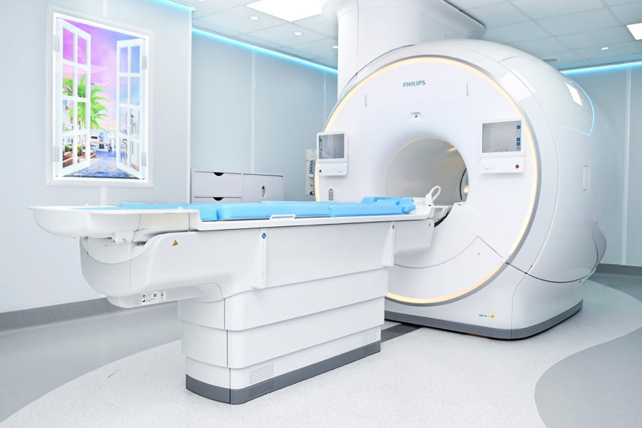
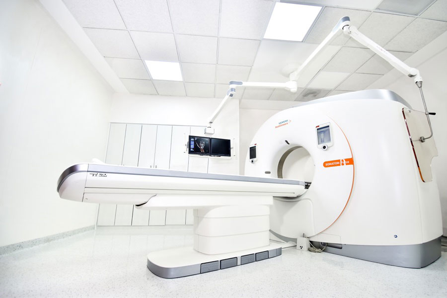
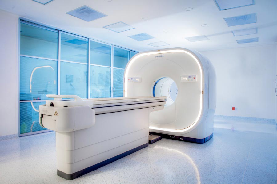
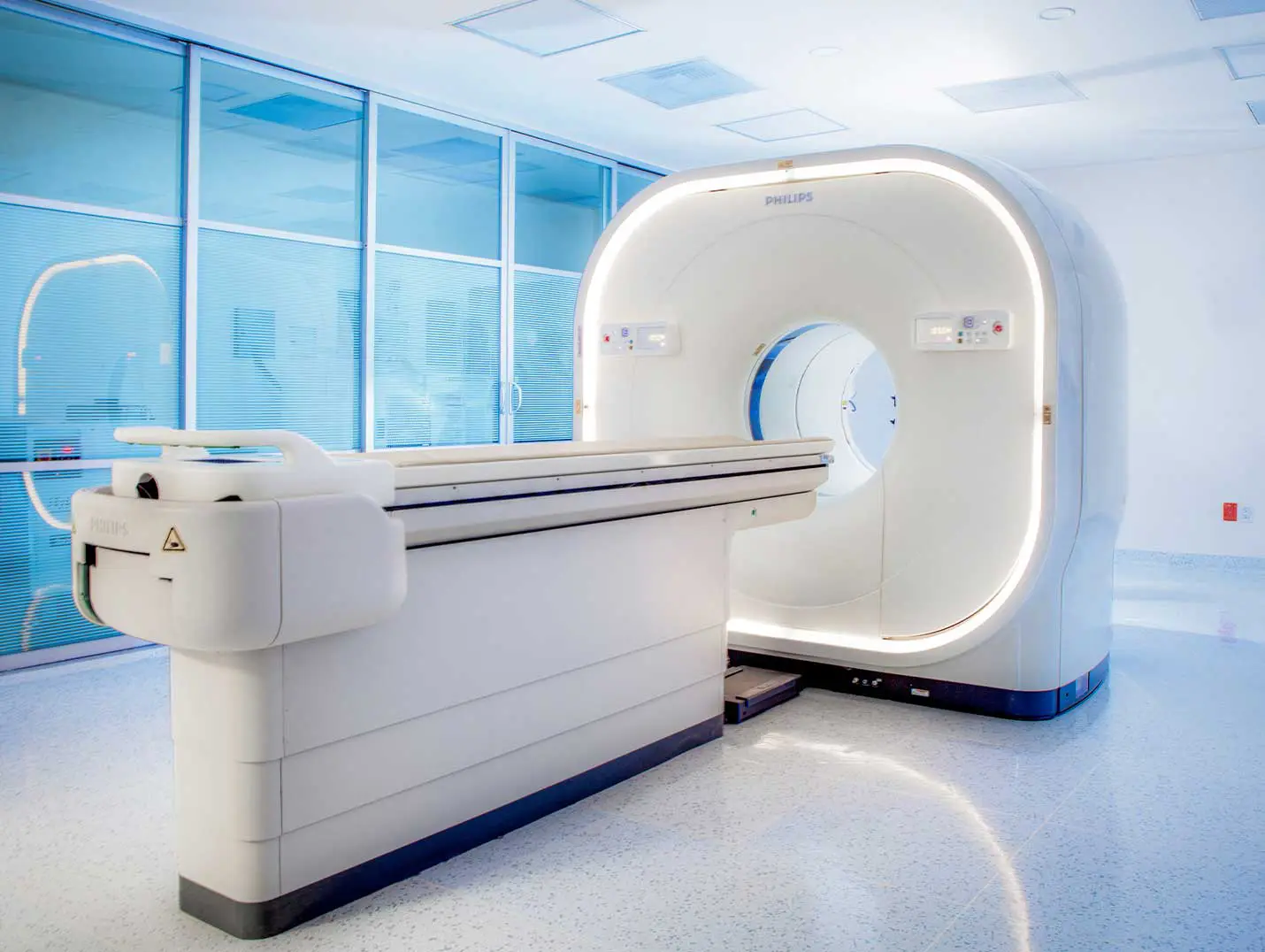
Positron emission tomography (PET-CT) is an imaging technique that combines computed tomography (CT) and positron emission tomography (PET) to produce detailed images of the inside of the body.
Positron emission tomography (PET-CT) is primarily used to detect and evaluate conditions such as cancer, heart disease, and neurological disorders. It is also used to assess response to treatment and plan surgeries.
During the procedure, the patient is injected with a small amount of a radioactive substance called a tracer. A series of images is then taken using a machine that combines computed tomography (CT) and positron emission tomography (PET).
Yes, positron emission tomography (PET-CT) is a safe procedure. However, because it uses radiation, there is a small risk of radiation exposure. The benefits of the procedure usually outweigh the associated risks.
Yes, before the procedure, you need to follow certain instructions, such as avoiding food and drink for several hours before the exam. It is also important to tell your doctor about any medications you are taking.